The detection of brain tumors typically involves a combination of medical history evaluation, physical examinations, and various medical imaging techniques. Here's an overview of the common methods used for brain tumor detection:
- Medical History and Physical Examination: The first step is often a thorough review of the patient's medical history, including symptoms, family history, and any relevant medical conditions. A neurological examination may be conducted to assess motor skills, sensory function, coordination, reflexes, and other neurological signs.
- Imaging Techniques:
- MRI (Magnetic Resonance Imaging): MRI scan is a powerful imaging technique that uses strong magnetic fields and radio waves to generate detailed images of the brain's structures. It can provide high-resolution images that help visualize the size, location, and characteristics of brain tumors.
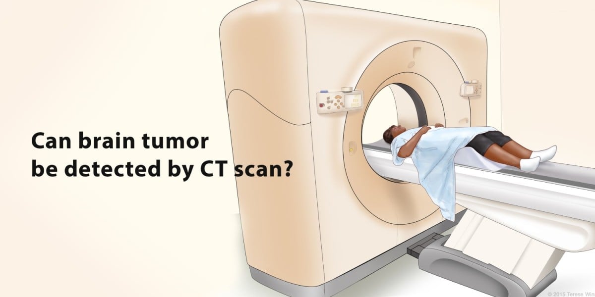
- CT (Computed Tomography) Scan: CT scans use X-rays to create cross-sectional images of the brain. While not as detailed as MRI for soft tissue, CT scans are useful for detecting abnormalities, including tumors.
- PET (Positron Emission Tomography) Scan: PET scans involve injecting a small amount of radioactive material into the body. This material accumulates in areas with high metabolic activity, such as tumors. A PET scan can provide information about the activity of a brain tumor.
- Functional MRI (fMRI): This type of MRI ct scan is used to map brain activity by measuring changes in blood flow. It can help determine if the tumor is affecting specific brain functions.
- Biopsy: In some cases, a biopsy may be necessary to definitively diagnose the type of brain tumor. During a biopsy, a small sample of the tumor tissue is extracted and examined under a microscope. This helps determine whether the tumor is benign (non-cancerous) or malignant (cancerous), as well as its specific characteristics.
- Cerebrospinal Fluid Analysis: In certain cases, particularly if a tumor is suspected to be affecting the spinal cord or certain areas of the brain, a sample of cerebrospinal fluid (CSF) may be collected and analyzed for abnormalities.
- Angiography: Angiography involves injecting a contrast dye into the blood vessels and taking X-ray images. This technique can help visualize the blood vessels in the brain and identify any abnormalities, including tumors that might be affecting blood flow.
- Genetic Testing: Genetic testing of tumor tissue can provide valuable information about the genetic mutations and markers present in the tumor. This information can help guide treatment decisions and provide insights into the tumor's behavior.
It's important to note that the specific approach to brain tumor detection can vary based on the patient's symptoms, medical history, and the suspected type of tumor. Medical professionals, including neurologists, neurosurgeons, and best radiologists in Gurgaon, work together to determine the most appropriate diagnostic methods for each individual case.